Uncovering Novel Mechanisms of Ferroptosis Defense and Triggering [Sep 9, 2025]


New Insights Reveal How Lipoproteins Protect Against Ferroptosis and EMCSs Initiate It
Ferroptosis is intensively studied as a therapeutic target in cancer and a pathogenic mechanism in neurodegeneration. One study reveals an unexpected role for lipoproteins, showing that beyond serving as nutrient sources or substrates for peroxidation, they also deliver antioxidant lipids such as vitamin E that protect cancer cells from ferroptosis1. Another introduces ER–mitochondria contact sites (EMCSs) as a newly recognized hub of PUFA-containing phospholipid peroxidation2, highlighting an additional mechanism that can initiate ferroptosis.

1. Glycosaminoglycan-mediated lipoprotein uptake protects cancer cells from ferroptosis (Nature, 2025)
Summary: Cancer cells actively increase the uptake of lipoproteins, lipid-carrying particles in the blood, through cell-surface glycosaminoglycans, sugar chains that act as binding sites and are elevated in tumours. This uptake represents a survival strategy, as the imported lipoproteins provide vitamin E (α-tocopherol) and other antioxidant lipids that block lipid peroxidation, suppress ferroptosis, and sustain tumour growth.

Highlighted technique: To investigate the impact of lipoproteins on ferroptosis, cancer cells were treated with GPX4 inhibitors to induce ferroptosis and cultured with lipoproteins or components, while lipid peroxidation was measured. These experiments showed that among lipoprotein-derived molecules, α-tocopherol is the principal factor suppressing lipid peroxidation and thereby preventing ferroptosis.

 Related technique  Lipid Peroxides Detection, Ferrous ion (Fe2+) Detection

2. Endoplasmic reticulum–mitochondria contacts are prime hotspots of phospholipid peroxidation driving ferroptosis (Nature Cell Biology, 2025)
Summary: The ER produces phospholipids, including polyunsaturated fatty acid (PUFA)-containing species, and supplies them to mitochondria at ER–mitochondria contact sites (EMCSs). Under ferroptosis-inducing conditions, EMCSs act as the initial hub where PUFA-containing phospholipids are oxidized. This local peroxidation triggers EMCS expansion, thereby increasing mitochondrial ROS and promoting mitochondrial fission, which together amplify ferroptosis.

Highlighted technique: To track the dynamics of lipid peroxides under ferroptosis-inducing conditions, cells were transfected with a fluorescently labeled ER marker and co-stained with mitochondrial and lipid peroxide probes. Time-lapse super-resolution live imaging with 3D rendering revealed that lipid peroxide signals first increased rapidly at EMCSs and subsequently redistributed to mitochondria. Furthermore, mitochondrial ROS measurements confirmed a marked increase following this redistribution.

 Related technique  Lipid Peroxides Detection, Mitochondrial ROS Detection

Related Techniques (click to open/close)
Target Kit & Probes
Ferroptosis Indicator: ferrous ion (Fe2+) FerroOrange(intracellular), Mito-FerroGreen(mitochondria)
Ferroptosis Indicator: lipid peroxidation Liperfluo(intracellular), MitoPeDPP(mitochondria)
Lipid Peroxidation Assay Lipid Peroxidation Probe -BDP 581/591 C11-
Cystine Uptake detection Cystine Uptake Assay Kit
Glutathione Quantification GSSG/GSH Quantification Kit
Mitochondrial superoxide detection MitoBright ROS Deep Red - Mitochondrial Superoxide Detection
Mitochondrial Staining MitoBright LT Green / Red / Deep Red
Total ROS detection Highly sensitive DCFH-DA or Photo-oxidation Resistant DCFH-DA
Apoptosis detection in multiple samples Annexin V Apoptosis Plate Assay Kit
Cell proliferation/ cytotoxicity assay Cell Counting Kit-8 and Cytotoxicity LDH Assay Kit-WST
Application Note (click to open/close)
  > When Lysosomes Go Neutral: Iron Loss Unveiled

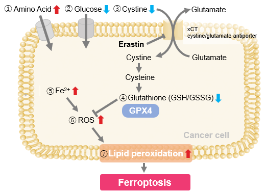
We investigated the transition of cellular metabolisms in A549 cells treated with erastin, a known ferroptosis inducer. Our results revealed the following.

Results
- The inhibition of cystine uptake by erastin led to a depletion of cysteine, which in turn increased the compensatory uptake of other amino acids.
- Glucose uptake, which typically promotes ferroptosis*, was found to decrease upon erastin treatment, suggesting a potential cellular self-defense mechanism.
- The depletion of cysteine resulted in a decrease in glutathione levels and an increase in Fe2+, ROS, and lipid peroxides, all of which are recognized markers of ferroptosis.

  Cell Line: A549
  Incubation Conditions: 100 μmol/l Erastin/MEM, 37℃, 3h
  *Reference: Xinxin Song, et al., Cell Reports, (2021)


Products in Use
① Amino Acid Uptake: Amino Acid Uptake Assay Kit
② Glucose Uptake: Glucose Uptake Assay Kit-Green
③ Cystine Uptake: Cystine Uptake Assay Kit
④ Intracellular glutathione: GSSG/GSH Quantification Kit
⑤ Intracellular labile Fe: FerroOrange
⑥ Intracellular total ROS: ROS Assay Kit -Highly Sensitive DCFH-DA-
⑦ Lipid Peroxides: Liperfluo


Product Classification

Product Classification