Previous Science Note
|
Polyunsaturated fatty acids (PUFAs) influence cell survival by altering membrane lipid composition, with recent studies showing that changes in PUFA levels and trafficking can either protect neurons from stress or increase cancer cell sensitivity to ferroptosis. This Science Note introduces three studies that uncover how PUFA-containing phospholipids regulate cell death through mitochondrial interactions, lipid remodeling, and tissue-specific lipid depletion. |
||||||||||||||||||||||||||||
|
Neuronal polyunsaturated fatty acids are protective in ALS/FTD (Nature Neuroscience, 2025) Summary: Amyotrophic lateral sclerosis (ALS) and frontotemporal dementia neurons are depleted of protective polyunsaturated fatty acids (PUFA). Replenishing these fatty acids through diet and by boosting their production inside the cells markedly prolonged the survival of both fly and human neuron models, suggesting a simple protective strategy. Highlighted technique: This study used iPSC-derived spinal neurons from ALS/FTD patients to test whether increasing lipid unsaturation could protect against neurotoxicity. Overexpressing desaturase enzymes raised PUFA levels and significantly reduced glutamate-induced neuronal death, showing a protective effect in this human disease model. Related technique Apoptosis Detection |
||||||||||||||||||||||||||||
|
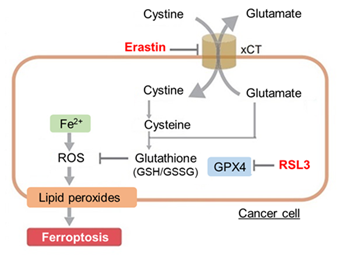
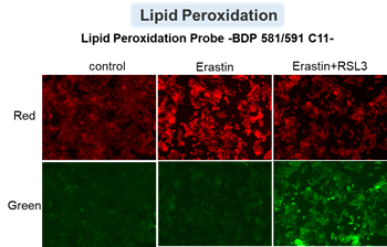
Phospholipids with two polyunsaturated fatty acyl tails promote ferroptosis (Cell, 2024) Summary: This study found that certain fatty acid treatments cause cells to build up PC-PUFA2, a lipid that binds mitochondria, generates reactive oxygen, and increases ferroptosis sensitivity. PC-PUFA2 was reduced in aging and Huntington disease brains, and mitochondrial antioxidants blocked its damaging effects. Highlighted technique: The researchers treated cancer cells with PC-PUFA2 lipids and measured ferroptosis sensitivity using lipid peroxidation and viability assays. They further quantified mitochondrial ROS and membrane potential changes, showing that PC-PUFA2 triggers ferroptosis through mitochondrial oxidative stress. Related technique Lipid Peroxide Detection (used in this article), OCR Assay |
||||||||||||||||||||||||||||
|
Summary: When cancer cells are starved of external lipids, they channel PUFAs into membrane phospholipids, which increases their sensitivity to ferroptosis. This suggests that reducing lipid availability around tumors could enhance PUFA driven ferroptosis based cancer therapies. Highlighted technique: The authors performed LC–MS/MS lipidomics using isotope-labeled PUFA to trace their incorporation into distinct phospholipid classes under lipid-starved conditions. They then assessed ferroptosis sensitivity by combining cell viability assays with lipid peroxidation staining after treatment with ferroptosis inducers. Related technique Lipid Peroxidation Assay, Cell Proliferation Assay |
||||||||||||||||||||||||||||
Related Techniques (click to open/close)
|
||||||||||||||||||||||||||||
Application Note (click to open/close)
|
||||||||||||||||||||||||||||
|
|
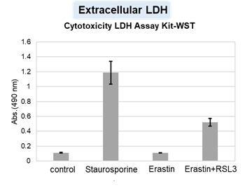
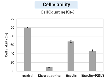
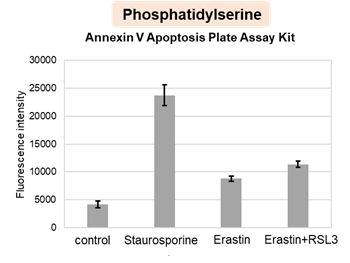
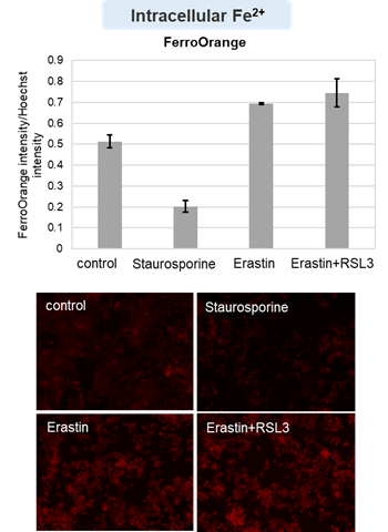
HepG2 cells treated with the apoptosis-inducing agent staurosporine or the ferroptosis-inducing agents Erastin and RSL3. After treatment, extracellular LDH, phosphatidylserine, cell viability, intracellular Fe2+ and lipid peroxidation were determined. |
||
  |
  |
 |
|