


Science Note / Mitophagy and Immunity
What Does Mitophagy Reveal About Immunity?
What can mitophagy reveal about inflammation and immune function? Cytokine levels alone cannot show whether a source of inflammation has changed or how well immune cells still work. One recent study showed that mitophagy reduced mitochondrial DNA outside mitochondria and limited inflammatory cell death. A separate human trial found changes in immune cell metabolism and improved responses to stimulation after treatment with a compound known to induce mitophagy. Together, these findings support measuring mitophagy and immune function alongside inflammatory signals.
← Previous Science NoteNew Insights from Recent Studies
Novel mitophagy activator TJ0113 restricts cytosolic mtDNA to suppress ZBP1-driven inflammation in acute lung injury
This study showed how DNA released from damaged mitochondria can trigger inflammatory cell death in acute lung injury. It also found that mitophagy can interrupt this process by removing damaged mitochondria before they release DNA.
View Summary & Techniques
This study identified how damaged mitochondria drive inflammation in acute lung injury. DNA leaking from them is detected by a sensor inside lung immune cells, activating pathways linked to two forms of inflammatory cell death, necroptosis and pyroptosis. Mitophagy removes damaged mitochondria, reducing DNA leakage and suppressing the responses that lead to both forms of cell death. These findings suggest that removing damaged mitochondria through mitophagy may help prevent inflammatory cell death by limiting the DNA signal that sets it in motion.
The researchers treated lung immune cells with a bacterial component to induce inflammation. After adding a mitophagy-promoting compound, they used live-cell imaging to assess the overlap between mitochondrial and lysosomal signals. They also measured mitochondrial membrane potential and mitochondrial ROS to examine whether changes in mitophagy were accompanied by changes in mitochondrial condition and oxidative stress.
Measuring mitochondrial membrane potential and mitochondrial ROS alongside mitophagy shows whether increased mitochondrial removal is accompanied by restored membrane potential and reduced oxidative stress.
Effect of the mitophagy inducer urolithin A on age-related immune decline: a randomized, placebo-controlled trial
In a placebo-controlled trial involving healthy middle-aged adults, urolithin A, a compound known to promote the removal of damaged mitochondria, increased certain T cells’ capacity to use fats and amino acids for energy. T cells collected from participants also produced more immune signaling molecules when stimulated in the lab, while monocytes became better at taking up bacterial particles.
View Summary & Techniques
In a placebo-controlled trial, four weeks of urolithin A increased certain T cells’ capacity to use fats and amino acids for energy. T cells collected from participants also responded more strongly to stimulation in the lab, while monocytes took up more bacterial particles. Together, these results show that an intervention using a compound known to induce mitophagy can change immune cell metabolism while improving the immune functions tested.
To examine mitochondrial responses to urolithin A, a compound known to induce mitophagy, the researchers treated human immune cells from separate healthy donors outside the body. Using flow cytometry, they detected early changes in mitochondrial membrane potential and lysosomal staining, followed by a decrease in mitochondrial mass. This sequence was consistent with mitochondrial removal in the treated cells.
Mtphagy Dye becomes brighter when labeled mitochondria reach acidic lysosomes, and this increase in fluorescence can be measured by flow cytometry. Combined with lysosome staining, it also allows their overlap to be observed by confocal microscopy.
Experimental Workflow
What Changes When Damaged Mitochondria Are Cleared?
Assess mitochondrial condition and delivery to lysosomes, then compare metabolism, inflammatory signals, and immune function.
Damaged Mitochondria
Mitochondrial DNA can appear outside mitochondria and trigger inflammatory responses.
- Measure mitochondrial membrane potential
- Measure mitochondrial superoxide
- Assess cytosolic mitochondrial DNA
Clearance
Track delivery of mitochondria to lysosomes and assess mitochondrial condition.
Metabolism and Immune Response
Compare metabolic activity, cytokines, and responses of immune cells after mitochondrial clearance.
- Measure oxygen consumption
- Measure cytosolic mtDNA and inflammatory cytokines
- Assess responses to stimulation and phagocytosis
Application Notes
Published experimental examplesThese examples illustrate complementary lysosomal and mitochondrial measurements. They use different models and treatments from the two studies above.
Application Note I
NAD+ Depletion and Autophagy-Lysosomal Pathway Response
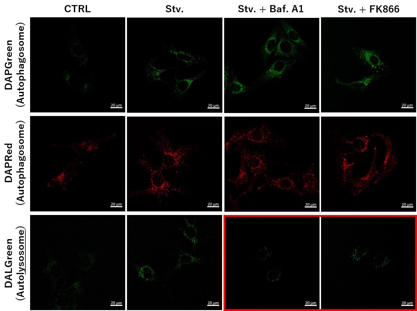
In HeLa cells, starvation increased signals from autophagosome and autolysosome probes. With FK866 or Bafilomycin A1 under starvation, DALGreen signal decreased, consistent with inhibition of progression toward autolysosomes associated with lysosomal deacidification.
Model: HeLa cells · Treatments: HBSS starvation, FK866, Bafilomycin A1 Readouts: DAPGreen, DAPRed, DALGreen
Application Note II
Simultaneous Detection of Lysosomal and Mitochondrial Dysfunction
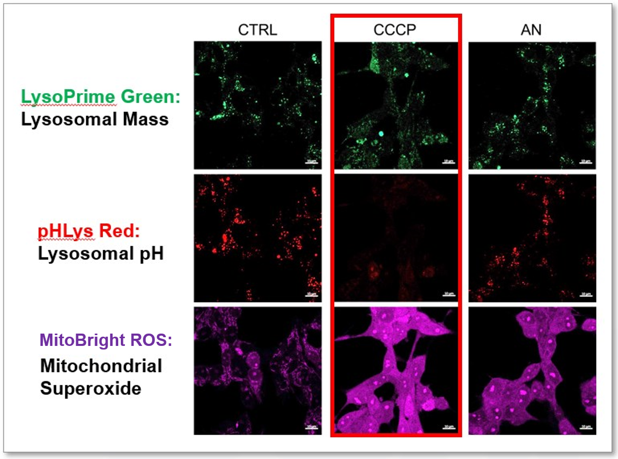
In HeLa cells treated with CCCP or antimycin, the example used MitoBright ROS Deep Red to detect mitochondrial superoxide and separate lysosomal dyes to assess lysosomal mass and pH. Co-staining showed lysosomal neutralization together with mitochondrial ROS induction after CCCP treatment.
Model: HeLa cells · Treatments: CCCP, antimycin
Readouts: MitoBright ROS Deep Red, LysoPrime Green, pHLys Red