Previous Science Note
| Autophagy and glycolysis are fundamental to maintaining cellular energy homeostasis, yet their imbalance can lead to dysfunction and disease. Recent studies reveal how impaired autophagy disrupts glucose metabolism and how excessive glycolytic flux can suppress autophagy.This Science Note introduces new insights into the dynamic crosstalk between these pathways and its impact on cellular homeostasis. | ||||||||||||||||||||||||
|
Autophagy regulator ATG5 preserves cerebellar function by safeguarding its glycolytic activity (Nature Metabolism, 2025) Highlighted technique: To assess changes in glucose uptake in autophagy-deficient Purkinje cells, the authors used cerebellar organotypic slice cultures from mice and evaluated glucose uptake capacity by fluorescent microscopy using a fluorescent analog of glucose. Related technique Autophagic Flux Assay, Highly Sensitive Fluorescent Glucose Analog |
||||||||||||||||||||||||
|
Weak neuronal glycolysis sustains cognition and organismal fitness (Nature Metabolism, 2024) Highlighted technique: In this study, a mouse model was generated by overexpressing PFKFB3, a glycolysis-promoting enzyme that is normally degraded in neurons, to investigate the importance of weak neuronal glycolysis. Tissues and cells derived from these mice were used to examine NAD⁺ levels, mitochondrial activity, and autophagy. Related technique NAD/NADH Assay, Glycolysis/OXPHOS Assay, Mitochondrial ROS Detection |
||||||||||||||||||||||||
|
Autophagy counters inflammation-driven glycolytic impairment in aging hematopoietic stem cells (Cell Stem Cell, 2024) Highlighted technique: In this study, HSCs were isolated from aged mice subjected to a fasting/refeeding regimen and transplanted into recipient mice to assess their regenerative capacity. FCM was used to detect donor-specific markers and quantify the expansion of donor-derived cells, allowing evaluation of the functional restoration of HSCs by the fasting/refeeding treatment. Related technique Autophagic Flux Assay, Extracellular OCR Assay, Glucose Uptake Assay |
||||||||||||||||||||||||
Related Techniques (click to open/close)
|
||||||||||||||||||||||||
Application Note I (click to open/close)
|
||||||||||||||||||||||||
|
|
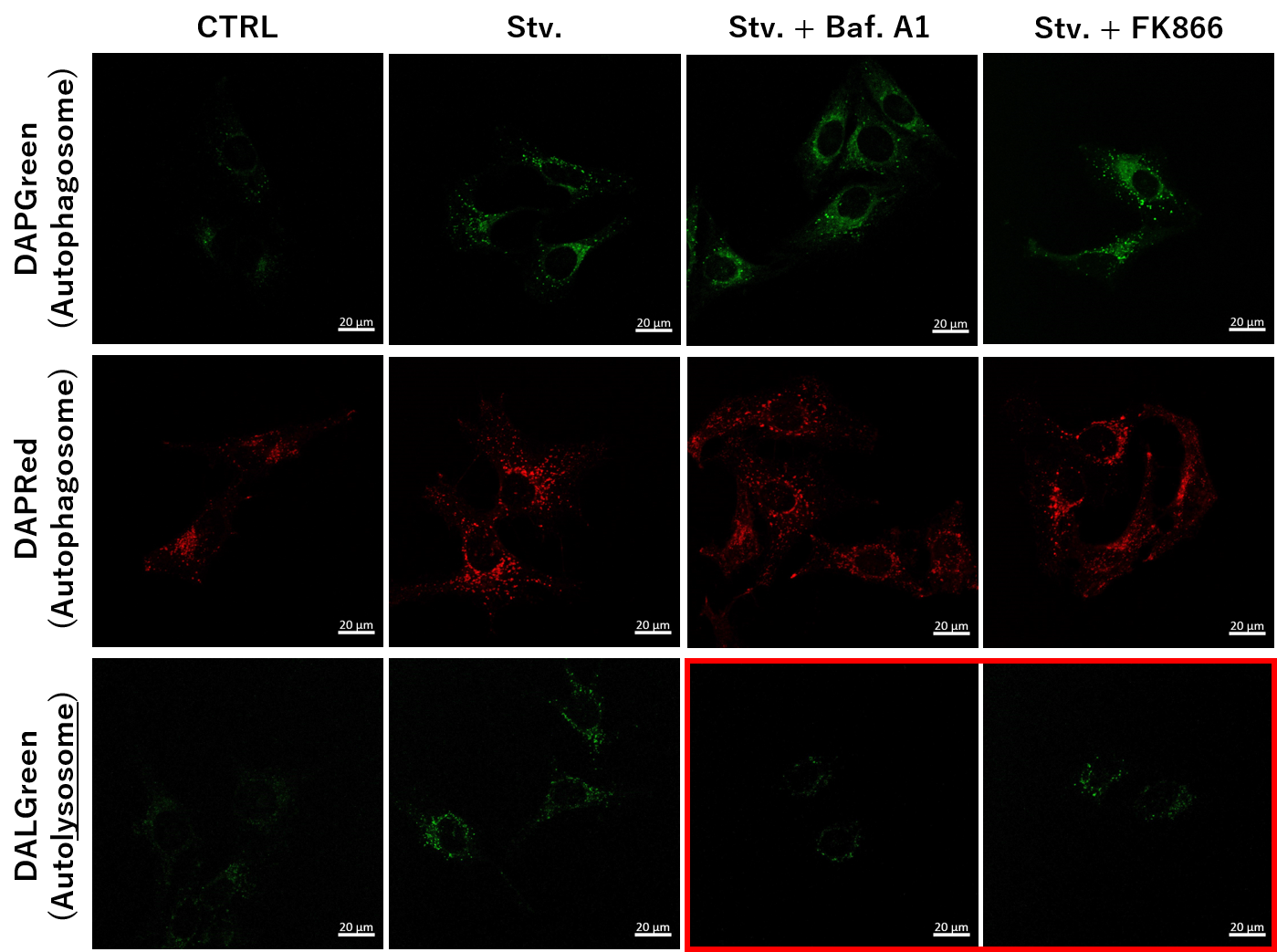
We determined how FK866-induced lysosomal deacidification affects the autophagy-lysosomal pathway. After staining with DAPGreen/DAPRed (for detecting autophagosome), or DALGreen (for detecting autolysosome), HeLa cells were starved in HBSS incubation and then treated with FK866 or Bafilomycin A1. Under the starvation condition, the fluorescent signals from all dyes increased, indicating the proceeding autophagy-lysosomal pathway. On the other hand, only DALGreen's signals were decreased in FK866 and Bafilomycin A1 treated cells with starvation conditions. These results clearly suggested that FK866 inhibits the autophagy-lysosomal pathway by NAD+ depletion-induced lysosomal deacidification. Nampt inhibitor, FK866 inhibits the progress of autophagosome to autolysosome by lysosomal deacidification. A recent finding shows that the dysfunctional condition of nicotinamide adenine dinucleotide (NAD+) biosynthetic enzyme, Nampt induces lysosomal deacidification1). In this section, we tried to determine how NAD+ depletion-induced lysosomal deacidification affects the autophagy-lysosomal pathway. 1) Mikako Yagi, et. al., EMBO J., 40(8), e105268 (2021) |
Application Note II (click to open/close)
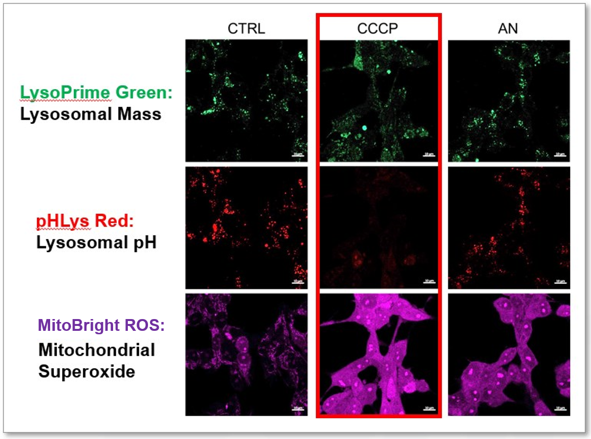
> Simultaneous Detection of Lysosomal and Mitochondrial Dysfunction
|
|
We tried the simultaneous detection of lysosomal and mitochondrial dysfunction in Hela cells treated with CCCP or Antimycin (AN). CCCP and AN are well-known inducers of mitochondrial ROS regarding loss of mitochondrial membrane potential. Recent research showed the result that CCCP induces not only mitochondrial ROS but also lysosomal neutralization. To detect mitochondrial ROS, HeLa cells were labeled by MitoBright ROS Deep Red - Mitochondrial Superoxide Detection, and the lysosomal mass and pH were detected separately with LysoPrime Green and pHLys Red. Co-staining with MitoBright ROS and Lysosomal dyes demonstrated that CCCP causes lysosomal neutralization and mitochondrial ROS induction at the same time. Products in Use
|