| [Apr. 23, 2024] | Previous Science Note |
|
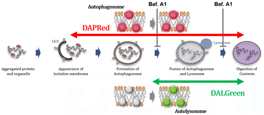
Autophagy is a cellular process that involves the degradation and recycling of cellular components through the formation of autophagosomes, which subsequently fuse with lysosomes for content degradation. In the context of cancer, autophagy plays a dual role; it can suppress tumor initiation by eliminating damaged organelles and proteins, but can also promote tumor survival and growth under metabolic stress by providing nutrients through the recycling of cellular components. |
|
|
Related Techniques |
|
|
|
|
|
|
|
|
Related Applications |
Analysis of autophagic flux without transfectionDetection PrincipleNampt inhibitor, FK866 inhibits the progress of autophagosome to autolysosome by lysosomal deacidification. A recent finding shows that the dysfunctional condition of nicotinamide adenine dinucleotide (NAD+) biosynthetic enzyme, Nampt induces lysosomal deacidification1). In this section, we tried to determine how NAD+ depletion-induced lysosomal deacidification affects the autophagy-lysosomal pathway. 1) Mikako Yagi, et. al., EMBO J., 40(8), e105268 (2021)
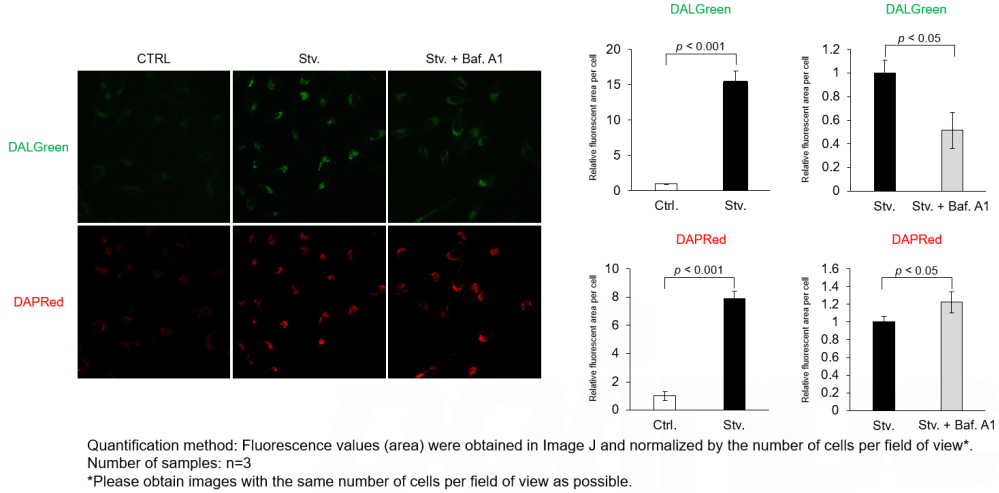
DALGreen and DAPRed labeled HeLa cells were used to evaluate changes in autophagic flux induced by the lysosomal acidification inhibitor bafilomycin A1 (Baf. A1). Compared to starvation conditions, the fluorescence signals of DALGreen were decreased under inhibited conditions of autolysosome formation by the addition of Baf. A1. In contrast, the fluorescence signals of DAPRed were increased under the same conditions, indicating that Baf. A1 led to the accumulation of autophagosome. Experimental Data
Experimental Conditions Procedure 1. HeLa cells were seeded (1.0 x 10⁴ cells/well) on a μ-slide 8 well plate (ibidi) and cultured overnight at 37°C in an incubator equilibrated with 95% air and 5% CO2. 2. After washing twice with MEM containing 10% fetal bovine serum, 200 μl of DALGreen/DAPRed working solution (DALGreen: 1 µmol/l, DAPRed: 0.2 µmol/l) and the cells were incubated at 37°C for 30 minutes. 3. The supernatant was discarded, and the cells were washed twice with MEM containing 10% fetal bovine serum. 4. Samples were prepared under the following conditions. 5. The stained cells were observed under a confocal fluorescence microscope. Products in Use |