

Previous Science Note
|
Extracellular ATP, released from dying or stressed cells, acts as a key damage-associated molecular pattern (DAMP) that signals immune cells and neighboring cells via purinergic receptors such as P2X7 and P2Y2. This Science Note introduces recent advances in our understanding of how extracellular ATP influences the immune system and the tumor microenvironment. |
||||||||||||||||||
|
Colon tumour cell death causes mTOR dependence by paracrine P2X4 stimulation (Nature, 2022) Colon tumor cells undergoing chemotherapy-induced cell death release extracellular ATP, which acts on neighboring tumor cells to activate mTOR signaling via P2X4 and promote their survival. Highlighted technique: This study used Lgr5-EGFP-DTR(+) mice, which allow selective induction of apoptosis in Lgr5-positive (stem) cells by diphtheria toxin. Colon tumor organoids derived from these mice enabled targeted ablation of Lgr5-positive tumor cells, allowing analysis of the impact of their death on neighboring Lgr5-negative cells. Related technique Extracellular ATP Assay, Annexin V Apoptosis Plate Assay |
||||||||||||||||||
|
To study the immune effects of ferroptosis cells, the researchers divided ferroptosis into initial, intermediate, and terminal phases, defined by lipid peroxidation, ATP release, and HMGB1 release with membrane rupture. Unlike apoptotic cells, ferroptosis cells fail to activate immunity through extracellular ATP and other signals, particularly in the initial phase, where they suppress dendritic cell maturation and phagocytosis. Highlighted technique: This study established a model to precisely control cell death by inducing GPX4 knockdown, which triggers ferroptosis, and temporarily blocking ferroptosis by adding ferrostatin-1. Removal of ferrostatin-1 triggers synchronised ferroptosis, allowing the process of ferroptosis to be precisely controlled. Related technique Intracellular Iron detection, Lipid Peroxide Detection |
||||||||||||||||||
|
In melanoma, extracellular ATP enhances the antitumor activity of CD8⁺ T cells by maintaining their mitochondrial function through the purinergic receptor P2RX7. Highlighted technique: Adoptive cell therapy (ACT) is a cancer treatment in which ex vivo activated immune cells are reinfused into the patient. As mitochondrial function is key to ACT efficacy, this study comprehensively evaluates T cell function using multiple parameters, including mitochondrial mass, mitochondrial ROS, oxygen consumption rate and extracellular acidification rate. Related technique Mitochondrial membrane potential detection, Mitochondrial superoxide detection |
||||||||||||||||||
Related Techniques (click to open/close)
|
||||||||||||||||||
Application Note I (click to open/close)
|
||||||||||||||||||
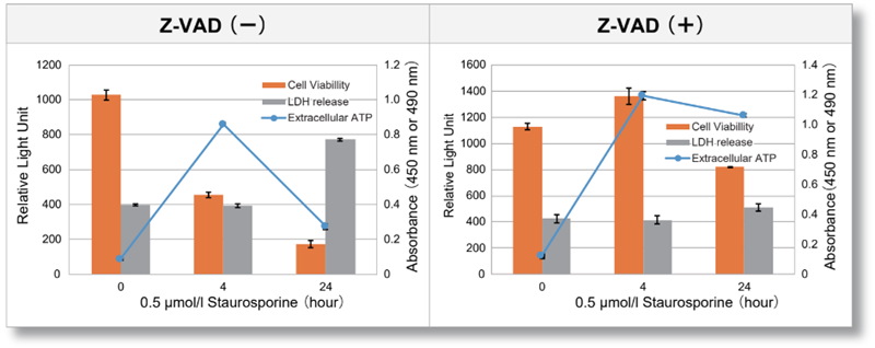
| Jurkat cells were treated with or without Z-VAD, an apoptosis inhibitor, and then treated with staurosporine. After treatment, changes in extracellular ATP release, cell viability and extracellular LDH release were assessed over time. The results showed that cell death was inhibited by Z-VAD, but extracellular ATP released during the initial phase of apoptosis increased over time. | |||
|
|
<Product in use> LDH release Cell viability |
||
Application Note II (click to open/close)
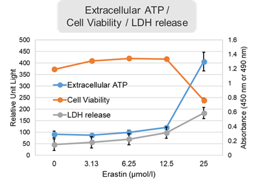
> Ferroptosis Induction and ATP Release Profile
|
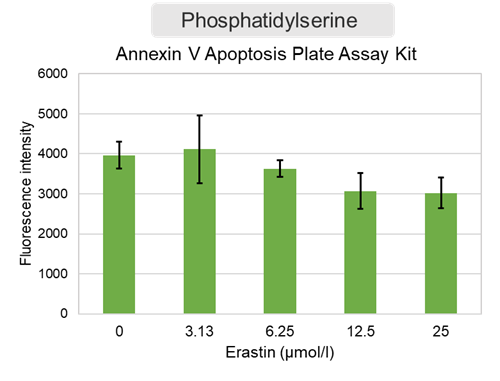
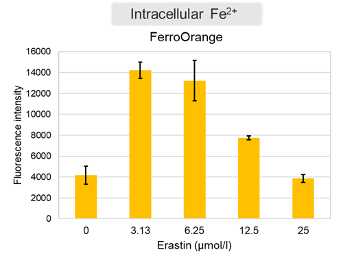
Changes in extracellular ATP release, cell viability, extracellular LDH release, phosphatidylserine, and intracellular Fe2+ were evaluated in HeLa cells treated with various concentrations of Erastin, a ferroptosis inducer, for 24 hours. The results showed that cell viability decreased and extracellular ATP release and extracellular LDH increased in cells treated with Erastin concentration of 25 μmol/l, indicating that cell death was induced under high concentration conditions. Interestingly, the increase in extracellular ATP in the early phase of stimulation, which was observed with the apoptosis inducer Staurosporine, was not observed with Erastin (See Experimental Example: Evaluation using Staurosporine-treated Cells.). Although the apoptosis-related marker phosphatidylserine was not significantly altered by Erastin treatment at any concentration. The amount of intracellular Fe2+, a ferroptosis-related marker, was significantly increased under the low-concentration treatment condition, indicating that it tends to increase before actual cell death occurs. |
<Product in use> Extracellular LDH: Cell viability: Cell Counting Kit-8 Phosphatidylserine: Intracellular Fe2+: FerroOrange |
||
 |
 |
 |
|


